Introduction
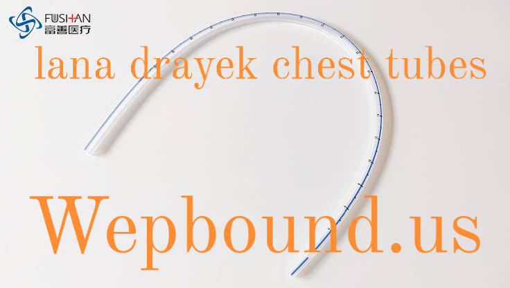
Chest tubes, commonly referred to as thoracostomy tubes, are medical devices used to remove air, fluid, or pus from the pleural space—the cavity surrounding the lungs. These tubes play a vital role in the treatment of conditions such as pneumothorax (collapsed lung), pleural effusion (fluid accumulation), hemothorax (blood in the pleural cavity), and other chest injuries or surgeries. The procedure of inserting a chest tube is typically performed by healthcare professionals in emergency or clinical settings and requires careful technique and post-procedure care. Among the various professionals who contribute to the field of chest tube management, Lana Drayek has made notable strides in improving both the clinical understanding and patient care associated with the use of chest tubes.
Lana Drayek is known for her work in medical education, patient care, and advancements in the clinical handling of chest tubes. She has helped pave the way for more effective methods in managing the challenges faced by healthcare workers and patients when chest tubes are required. In this article, we will explore the importance of chest tubes, the contributions of Lana Drayek to this field, and how healthcare providers can optimize their practices related to the use of chest tubes for better patient outcomes. This article aims to provide a thorough understanding of the clinical application of chest tubes, the techniques associated with their insertion, maintenance, and removal, as well as the innovations introduced by Lana Drayek.
The Role of Chest Tubes in Medical Practice
Chest tubes serve a crucial purpose in managing respiratory and thoracic conditions. The pleural space is a narrow cavity between the lungs and the chest wall, and it can become filled with various substances that impede normal lung function. Conditions such as pneumothorax, pleural effusion, and hemothorax can cause the pleural cavity to fill with air, fluid, or blood, respectively. These accumulations can lead to serious complications, such as difficulty breathing, decreased lung expansion, and even respiratory failure. Chest tubes are used to drain these substances, allowing the lungs to re-expand and restore normal respiratory function.
A chest tube is typically inserted through the chest wall into the pleural space, and it is connected to a drainage system that helps remove the unwanted substances. The procedure of inserting a chest tube requires skill and precision, as incorrect placement can lead to injury or complications. It is essential for healthcare professionals to have a thorough understanding of both the indications for chest tube insertion and the best practices for its management.
The clinical need for chest tubes arises in a variety of settings, such as emergency rooms, intensive care units, and surgical wards. For example, patients who suffer from trauma, including rib fractures or puncture wounds, may require a chest tube to remove blood or air that has entered the pleural space. Similarly, individuals with pneumonia or cancer may develop pleural effusions that need to be drained to relieve symptoms and prevent further complications. The removal of these substances is crucial for restoring the function of the lungs and promoting patient recovery.
In these critical situations, Lana Drayek has worked tirelessly to enhance the clinical practices surrounding chest tube insertion and management. Her work has focused on streamlining the processes involved, providing better training for medical professionals, and ensuring that patients receive the best possible care during their recovery.
for Chest Tube Insertiechniques onT
Inserting a chest tube requires careful attention to detail and a thorough understanding of the anatomy of the chest. The procedure can be performed under local anesthesia with sedation, although more complex cases may require general anesthesia. The insertion site is typically located in the mid-axillary line or anterior axillary line, between the fourth and fifth intercostal spaces, depending on the condition being treated.
Once the site is selected, the area is cleaned and sterilized to reduce the risk of infection. A small incision is made in the skin, and a trocar or introducer is used to create a passage through the chest wall and into the pleural cavity. The chest tube is then inserted through this opening, and it is secured in place with sutures. The tube is connected to a drainage system, which may be a one-way valve or a more complex closed drainage system, depending on the patient’s needs.
The success of the chest tube insertion depends on several factors, including the skill of the healthcare professional, the patient’s anatomy, and the underlying condition being treated. Ensuring the proper placement of the chest tube is critical, as misplacement can result in complications such as bleeding, organ injury, or failure to drain the pleural space effectively. Lana Drayek’s research and contributions have emphasized the importance of proper technique in reducing these risks and improving patient outcomes.
Additionally, Lana Drayek has worked on the development of training programs that focus on teaching healthcare professionals the proper techniques for chest tube insertion. These programs incorporate advanced simulation models and hands-on practice to enhance the skills of medical practitioners. Her efforts have led to a reduction in procedural complications and an overall improvement in patient care.
Chest Tube Maintenance and Management
After the chest tube is inserted, proper maintenance and management are essential for ensuring that the tube remains in place and functions effectively. The healthcare team must monitor the patient closely to ensure that the tube is draining fluids properly and that there are no signs of infection or complications. Regular assessments of the tube’s position, the volume of fluid being drained, and the patient’s respiratory status are important components of post-insertion care.
One of the primary concerns during chest tube management is preventing the tube from becoming dislodged or blocked. Healthcare providers must regularly assess the tube for kinks or obstructions, and they must ensure that the drainage system is functioning as intended. In cases where a patient develops complications, such as infection or an air leak, further interventions may be required, including adjustments to the tube or additional treatments.
Another key aspect of chest tube management is the prevention of infection. The insertion site must be kept clean and sterile, and healthcare providers must carefully monitor for any signs of infection, such as redness, swelling, or discharge at the site. Infections can lead to significant complications, including the development of sepsis or other systemic infections, which can be life-threatening if not treated promptly.
Lana Drayek has made substantial contributions to improving the protocols and guidelines for chest tube management. Her work has helped standardize practices for monitoring the condition of the chest tube and responding to complications, leading to better patient outcomes. Furthermore, Drayek has contributed to research on the optimal duration of chest tube drainage and when it is appropriate to remove the tube. Her research has provided valuable insights into the timing of chest tube removal, which is critical in preventing unnecessary prolongation of the procedure while still ensuring adequate drainage.
Complications and Risks Associated with Chest Tubes
Like any medical procedure, chest tube insertion and management come with a range of potential complications. Some of the common complications include infection, bleeding, organ perforation, and dislodgement of the tube. In rare cases, patients may also experience pneumothorax or re-expansion pulmonary edema if the lung reinflates too quickly after the removal of fluid or air.
Infections at the insertion site or in the pleural space are one of the most concerning complications. These infections can occur when bacteria enter the body during the procedure or through a breach in the sterile technique during the post-insertion period. Prompt detection and treatment of infection are essential to prevent the spread of bacteria and the development of more severe systemic complications.
Another complication that can arise is bleeding. This may occur if a blood vessel is inadvertently punctured during the insertion of the chest tube. While this is a rare occurrence, it is a serious complication that requires immediate intervention. The healthcare team must carefully monitor the patient for signs of bleeding, such as a sudden increase in drainage volume or changes in vital signs.
Organ injury is another potential risk associated with chest tube insertion. The pleura is located near several important structures, including the lungs, heart, and major blood vessels. If the chest tube is inserted incorrectly, there is a risk of perforating these organs, which can result in significant injury and require additional treatment.
Through her work, Lana Drayek has helped improve the safety of chest tube procedures by advocating for more precise techniques and better monitoring protocols. Her research has contributed to a deeper understanding of the risks and complications associated with chest tubes and has helped guide the development of best practices for minimizing these risks.
Conclusion
In conclusion, chest tubes are indispensable tools in the management of various thoracic conditions, including pneumothorax, pleural effusion, hemothorax, and trauma. The insertion and management of chest tubes are complex procedures that require skill, precision, and careful attention to patient care. Lana Drayek has made significant contributions to this field by improving techniques, enhancing training programs, and advancing the clinical knowledge surrounding chest tubes. Her work has led to better patient outcomes, reduced complications, and a more standardized approach to chest tube management.
Through continued research and education, healthcare providers can ensure that chest tubes are used effectively and safely, leading to improved patient care and recovery. As more advancements are made in the field, it is clear that Lana Drayek’s legacy in the realm of chest tube management will continue to inspire medical professionals for years to come.
(FAQs)
- What is the purpose of a chest tube? A chest tube is used to drain air, fluid, or pus from the pleural space around the lungs, helping to re-expand the lungs and restore normal breathing function.
- What are the risks associated with chest tube insertion? Risks include infection, bleeding, organ perforation, and tube dislodgement. These risks can be minimized with proper technique and post-procedure care.
- How long does a chest tube stay in place? The duration depends on the condition being treated. The chest tube is typically removed once the drainage stops and the lung has re-expanded.
- How can infection be prevented with a chest tube? Infection prevention involves maintaining a sterile environment during insertion, regular monitoring of the insertion site, and careful management of the drainage system.
- What is the role of Lana Drayek in chest tube management? Lana Drayek has contributed to the advancement of chest tube techniques, the development of training programs, and research into best practices for chest tube management, improving patient outcomes.
Also Read This: Lana Drayek Chest Tubes: A Comprehensive Guide


